
At Vista Eye Specialists, our talented eye doctors focus on restoring and maintaining the health of your eyes with the latest eye treatments. We stay up to date on all advances in medical and surgical therapies so we can offer you options in your eye care. Our team undergoes regular training to keep our knowledge and techniques current. Whenever possible, we incorporate leading-edge technology to make your treatment safer, quicker and more precise.
Our vision expertise includes:
- Contact Lens Fitting & Exam
- Eyelid Surgery / Blepharoplasty
- Pterygium Surgery
- Glaucoma
- Macular Degeneration
- Diabetic Eye Exam
- Dry Eye
- Lesion / Skin Growth Removal
- Comprehensive Eye Exams
CONTACT LENS FITTING & EXAM
When you wear contacts lenses, a precise fitting and comprehensive exam are essential to the clarity of your vision, as well as to your safety and satisfaction.
As trusted eye doctors serving Fredericksburg, Culpeper and surrounding communities, our doctors are focused on ensuring sharp vision and a comfortable fit for patients who rely on contact lenses, including conventional daily wear varieties, disposables, planned replacements, soft torics, rigid gas permeables, multifocal lenses, and scleral lenses.
EYELID SURGERY / BLEPHAROPLASTY
Vista Eye offers the state-of-the-art eyelid surgery to minimize the signs of aging around the eyes.
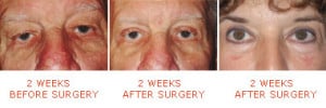
Eyelid surgery (blepharoplasty) is a surgery that reshapes the upper eyelid and/or lower eyelid by the removal of excess tissue. It can reduce sagging or puffiness, folds, and under eye “bags” that create a tired or stressed appearance. Blepharoplasty may be recommended for you if your vision is impeded by skin hanging down around your eyes. With blepharoplasty surgery, Dr. Jani can remove the excess skin and underlying fat.
The blepharoplasty procedure is performed by making small external incisions along the natural lines of the eyelids, such as the creases of the upper lids and below the lashes of the lower lids. The skin is then separated from the underlying fatty tissue. Finally excess fat and excess skin are removed and the incisions closed. The procedure typically takes 30 minutes to two hours and the initial recovery takes one to two weeks for the swelling and bruising to resolve, while final results can be seen after several weeks to a few months.
Benefits of Blepharoplasty
More than any other facial feature, the eyes have a tremendous impact on your appearance, and are often the first feature to show signs of aging. Vista Eye offers state-of-the-art eyelid surgery, also known as blepharoplasty, to minimize the signs of aging around the eyes. With blepharoplasty surgery, Dr. Jani can remove excess skin and fat that causes puffiness, droopiness and otherwise tired or sad-looking eyes.
Eyelid surgery can also treat excess tissue folds that may droop over the upper lash line into the line of sight. This is a great option if your vision is impeded due to skin hanging down around your eyes.
Blepharoplasty: Overview
ARE YOU A GOOD CANDIDATE FOR EYELID SURGERY?
 Generally speaking, you may be a good candidate for blepharoplasty if you:
- Are bothered by puffy or saggy eyelids
- Have folds of skin that droop over the upper lash line
- Find your vision is impaired because your upper eyelids droop so low
- Have trouble wearing glasses or contact lenses because of excess eyelid tissue
- Have under-eye bags or puffiness that makes you look tired or stressed
Dr. Jani will want to evaluate you in person to examine your eyelid anatomy and determine whether you are a suitable candidate for surgery. You should be in good health without any serious medical conditions that could affect your recovery from surgery, or any serious eye conditions that could jeopardize your surgery or outcomes. You should not smoke, or be able to quit several weeks before and after surgery. You should have realistic goals of treatment, which you can discuss at length with Dr. Jani during your consultation.
WHAT HAPPENS DURING EYELID SURGERY?
 Eyelid surgery is normally performed on an outpatient basis at a facility in Fredericksburg, so you can recover at home after the operation. Anesthesia will be used so you don’t feel any discomfort during the procedure.
To begin, Dr. Jani will create the surgical incisions along the natural lines and creases of your eyelids, such as the creases of the upper eyelids or below the lashes of the lower eyelids. The incision pattern is designed so that any scarring will be concealed within the natural structures of the eyelids. Sometimes he uses the transconjunctival incision on the inside of the lower lid.
Dr. Jani will then separate the skin from the underlying tissue and remove or reposition the tissue and fat as needed. He can also trim loose skin and tighten the remaining lid to create firmer and smoother eyelids. When he is finished, he closes the incisions with small sutures or stitches. The procedure typically takes 40 to 90 minutes.
RECOVERING FROM EYELID SURGERY
Initially after eyelid surgery, you can expect your eyes and eye area to be mildly swollen and bruised. Your eyelids may feel numb or tight. The majority of these side effects should subside within one to two weeks, at which point you can conceal any remaining bruising with makeup. Vista Eye will give you specific instructions on how to care for your eyelids as they heal.
You may notice an improvement in your eyelids immediately after surgery, with the results continuing to improve over the next few weeks and months.
Eyelid surgery will not stop the aging process, and the results will not last forever, but your eyes will feel and look younger.
PTERYGIUM SURGERY
Pterygium is a dense growth of tissue that grows across the cornea. Although the exact cause is unknown, it usually describes the condition of hazy vision and eye irritation usually caused by over-exposure to the sun’s harmful UV rays or environmental irritants like dust and wind. When a pterygium is large, it can become inflamed and painful. It can also cause blurry vision because it interferes with the focusing ability of the eye. In extreme cases, the pterygium can grow completely across the front of the eyeball and lead to significant loss of vision.
Pterygium: Overview
How Pterygium Surgery Works
At Vista Eye, the removal of pterygium may be a simple, in-office procedure that only takes about 10 minutes to perform. The surgeon will administer a local anesthetic to help the patient remain comfortable throughout the procedure. The pterygium will be removed in a no-stitch surgical procedure, after which the patient may experience some redness and irritation. To soothe the discomfort, your eye surgeon will prescribe an antibiotic ointment and patch the eye, which should be worn overnight. The patient will be required to return to our office the day after his or her procedure for follow-up treatment. There is very little downtime associated with pterygium removal, with most patients able to return to work and normal activities the next day.
Why Get Treatment
If left untreated, the pterygium growth will most likely continue to the point that it will take over a significant part of the cornea, at which time the patient’s vision could be almost completely blocked. Success rates with pterygium removal are typically very high, although there is a chance the pterygium may grow back following removal. If you are suffering from redness, irritation and blurry vision and are unable to find relief from glasses, contacts and artificial tears, you may be a candidate for pterygium removal.
GLAUCOMA TREATMENT
Glaucoma is a leading cause of blindness in the United States. Glaucoma is a disease of the optic nerve. When the pressure inside the eye rises, the optic nerve is damaged. The optic nerve connects the eye to brain, so that this affects the transmission of information from your eyes to your brain, resulting in eventual vision loss. The condition often develops over many years without causing pain or other noticeable symptoms – so you may not experience vision loss until the disease has progressed. If left untreated, glaucoma may cause irreversible optic nerve damage and blindness. There is no better reason to undergo regularly-scheduled, comprehensive eye exams. With annual eye exams, vision loss from glaucoma can be prevents. Visit our glaucoma page to learn more.
MACULAR DEGENERATION TREATMENT
Macular degeneration is a medical condition usually of older adults that results in a loss of vision in the center of the visual field (the macula) because of damage to the retina. The macula is a part of the retina in the back of the eye that ensures that our central vision is clear and sharp. The macula is used for reading, driving, recognizing faces, and watching television.
Age-related macular degeneration (ARMD) occurs when the arteries that nourish the retina harden. Deprived of nutrients, the retinal tissues begins to weaken and die, causing vision loss. Patients may experience anything from a blurry, gray or distorted area to a blind spot in the center of their vision.
Macular degeneration is the leading cause of legal blindness in people over age 55. Macular degeneration doesn’t cause total blindness because it doesn’t affect the peripheral vision. Symptoms include a gradual loss of the ability to see objects clearly, a gradual loss of color vision, distorted or blurry vision, and dark/empty areas appearing in the center of your vision. Possible risk factors include genetics, age, diet, smoking and sunlight exposure.
Regular eye exams are highly recommended to detect macular degeneration early and prevent permanent vision loss. Recent developments in ophthalmology allow doctors to treat many patients with early-stage ARMD with the help of lasers and medications.
DIABETIC EYE EXAM
Diabetic retinopathy is a complication of diabetes that weakens the blood vessels that supply nourishment to the retina (the light-sensitive lining in the back of the eye where vision is focused). These weak vessels can leak, swell or develop thin branches, causing a loss of vision. Changes to your vision may not be noticeable at first. But in its advanced stages, the disease can cause blurred or cloudy vision, floaters and blind spots and eventually, blindness. This damage is irreversible.
Diabetic retinopathy is the most common diabetic eye complication and a leading cause of blindness in American adults. Macular edema, which is leaking fluid that causes blurred vision, often occurs with diabetic retinopathy.
Fortunately, diabetic retinopathy is preventable. Your risk is reduced if you follow your prescribed diet and medications, exercise regularly, control your blood pressure, and avoid alcohol and cigarettes. Regular eye exams are an integral part of making sure your eyes are healthy.
Diabetic retinopathy can be detected through a comprehensive dilated eye exam. Although damage caused by diabetic retinopathy cannot be corrected, patients diagnosed with the condition can be treated to slow its progression and prevent further vision loss. Treatment modalities include laser, medications, and surgical procedures.
DRY EYE TREATMENT
Dry Eye Syndrome (Keratitis Sicca) occurs when the eyes aren’t sufficiently moisturized, leading to itching, redness, and pain from dry spots on the surface of the eye. The eyes may become dry and irritated, characterized by a scratchy, gritty feeling, because the tear glands don’t produce enough tears or because the tears themselves have a chemical imbalance, which is caused by abnormal tear composition resulting in rapid evaporation or premature destruction of the tears.
Your eyes require a constant layer of tears to lubricate, nourish, protect and cleanse your eyes. With chronic dry eye the underlying changes of the health of the tear-producing glands and their inability to produce the correct amount of tears results in a tear film that does not provide proper nourishment or protection. This may lead to the irritating symptoms of dry eye. Dry eyes can occur as a result of aging, environmental factors, medical conditions, or medication side effects.
Dry eye is not only painful, but it can also damage the eye’s surface and impair vision.
Fortunately, many treatment options are available. Non-surgical treatments for dry eye include increasing humidity at home or work and use of artificial tears or moisturizing ointment. Many individuals benefit topical drops that increase tear production from the surface glands on the eye. If these methods fail, our eye surgeon can insert small punctal plugs in the corners of the eyes to limit tear drainage or close the drainage tubes in the eyes in a simple office procedure. The plugs help retain tears and give many patients relief from the symptoms of dry eyes. In addition, eyelid surgery is also a solution if an eyelid condition is causing your dry eyes.
Do You Have Dry Eye?
 Every time you blink, tears are spread across the front surface of the eye. Your eyes depend on a flow of tears to provide constant moisture and lubrication to maintain eye health and comfort. When the volume or consistency of the tear system becomes imbalanced, you might experience dry eyes.
When tears do not adequately lubricate the eye, a person may endure:
- Sensitivity to light
- Blurred vision
- Burning sensation
- Discomfort in windy or dry conditions
- A gritty sensation
- Redness
- Eye Fatigue
Although the symptoms often start as minor inconveniences, the effects of dry eye are progressive and can worsen over time, potentially leading to severe discomfort or blurred vision.
Not Enough Tears?
Tears are necessary for your overall eye health.
 Dry Eye occurs when the eyes do not produce sufficient tears properly or the tears evaporate too quickly. Without proper treatment, inadequate tear production can make daily activities frustrating, affecting your personal and professional life, from reading or watching TV, to driving or working on the computer.
The most common form of Dry Eye is the Evaporative form, which affects 8 out of every 10 dry eye cases (86%). This indicates a shortage of oil on the surface of your tears, leading them to evaporate faster than normal. This shortage of tear oil is caused by a blockage in your eyelid (Meibomian) glands and is referred to as Meibomian Gland Dysfunction (MGD).
Dry Eye Questionnaire Form
LESION / SKIN GROWTH REMOVAL
At Vista Eye, our doctors can help improve your appearance by removing small lesions and/or skin growths on the face and neck that may be hereditary or have developed through the natural aging process over the years. Our experienced surgeon can perform a simple office procedure to remove these lesions and skin growths for an immediate improvement to your complexion.
Frequently Asked Questions About Eye Care
What does “20/20 vision” mean?
If an eye doctor tells you that you have 20/20 vision, it means that you meet the standard of visual acuity at a distance. This is measured through a test in which you are asked to read letters of a certain size on a chart simulated as 20 feet away.
Visual acuity at a distance is only one measure of how well you can see. Other important visual functions include peripheral vision, near vision, contrast sensitivity, color vision and depth perception.
Are there contacts that offer clear close and distance vision?
Yes. Multifocal contact lenses offer good vision at near and far distances. There is a slight adjustment period at first, but many patients like these lenses and appreciate the flexibility they offer.
Sometimes I see tiny spots or squiggles that appear to float in front of my eyes. Are these any cause for concern?
These visual aberrations, called “floaters,” are usually harmless. Reasons to be concerned are if you see a sudden increase in the number of floaters, if the floaters themselves become very large and interfere with your vision or if you see flashing lights in your peripheral vision. In those scenarios, you would need to be evaluated by an eye doctor to rule out any problems with your retina.
Can prolonged screen time hurt my eyes?
Prolonged periods of time spent looking at a computer, tablet or smartphone screen can lead to tired or dry eyes. Your digital screen can strain your eyes and cause them to become fatigued. Staring at a screen also tends to slow down your natural blink rate, and when you do not blink as much as you should, your tear film does not spread over your ocular surface and your eyes can become dry and irritated.
Can exposure to ultraviolet light hurt my eyes?
Yes. Excessive exposure to ultraviolet light (UV) can cause short-term and long-term damage. UV exposure has been implicated as a risk factor for diseases such as cataracts and macular degeneration; and excessive UV exposure can cause skin cancer of the eyelids.
The best way to protect your eyes from the sun is to wear sunglasses that block 99 to 100 percent of both UV-A and UV-B radiation. Adding a broad-brimmed hat is also helpful. And, you should have your eyes examined by a doctor regularly to detect any signs of damage as early as possible.
What is the difference between an ophthalmologist and optometrist?
Ophthalmologists and optometrists are two different kinds of doctors that diagnose and treat eye conditions. Ophthalmologists attend medical school and have the designation of MD. Optometrists attend optometry school and have the designation of OD. Both are qualified to perform eye exams, prescribe glasses and contact lenses and manage diseases and disorders affecting the visual system, such as glaucoma, dry eye, and macular degeneration.
The primary difference between an ophthalmologist and optometrist is that an ophthalmologist is a medical doctor and is trained and experienced in eye surgery procedures such as LASIK and cataract surgery.
Schedule an Eye Consultation
If you are considering cosmetic procedures, schedule a consultation with Dr. Jani. He will discuss with you different treatment options and create a custom plan to help you achieve your goals and desired appearance. Please contact Vista Eye for more information on cosmetic procedures, or to schedule an appointment today.