

At Vista Eye Specialists, our experienced doctors are committed to delivering comprehensive glaucoma care for patients in Fredericksburg and Culpeper, Virginia. With consistent monitoring and early intervention, glaucoma can be managed effectively, protecting your vision for years to come.
Table of Contents
Understanding Glaucoma
Glaucoma is a leading cause of blindness in the United States. It’s a disease of the optic nerve. When the pressure inside the eye rises, the optic nerve is damaged. Because the optic nerve connects the eye to the brain, glaucoma affects the transmission of information from the eye to the brain, resulting in eventual vision loss.
Glaucoma often develops over many years without causing pain or other noticeable symptoms, so you may not experience vision loss until the disease has progressed. Once diagnosed, glaucoma may cause irreversible optic nerve damage and may lead to blindness if not treated. Early detection with a comprehensive eye exam is recommended to help prevent vision loss from glaucoma.
Types of Glaucoma
There are two primary types of glaucoma: open-angle glaucoma and angle-closure glaucoma. The “angle” refers to the drainage angle, located at the juncture of the iris and the pupil that controls the outflow of fluid. The eye continually produces a clear fluid, and in a healthy eye this fluid drains out of the eye through the drainage angle to keep the pressure inside the eye steady.

Symptoms of Glaucoma
In cases of open-angle glaucoma, there are often no symptoms until the late stages of the disease because the increase in fluid buildup and intraocular pressure happens slowly over time.
In the case of an angle-closure attack, symptoms do occur, and may include:
- Blurred vision
- Headaches
- Loss of peripheral vision
- Halo effects around lights
- Painful and / or reddened eyes
- Nausea and / or vomiting
Exams to Detect Glaucoma
During your exam, your eye pressure, the condition of the optic nerve, and your peripheral vision will be measured. If signs of glaucoma exist, more tests will be conducted. Our doctors at Vista Eye Specialists use an optical coherence tomography (OCT) to measure the thickness of the retinal nerve fiber layer, which is the tissue directly affected by glaucoma.
Thin nerve fiber readings may indicate the onset of glaucoma and the need for further testing. Dr. Jani will create a customized glaucoma treatment plan to help preserve and maintain the best possible vision for you. Regular eye exams help to monitor the changes in your eyesight and to determine whether you may develop glaucoma.
Glaucoma Risk Factors
People at high risk for glaucoma include those who are:
- Very nearsighted people (high myopia)
- People over the age of 40
- People with family history of glaucoma
- People with systemic diseases such as diabetes, anemia, or hardening of the arteries
- African-American or Hispanic/Latino
- Long-term steroid users
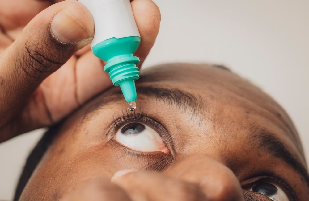
Treatment Options
When glaucoma is detected early, it can usually be controlled and further vision loss can be prevented. Treatments include:
iStent
In some MIGS procedures, tiny drainage tubes or “stents” are implanted in the eye to send the fluid into a reservoir. For example, the iStent is a microscopic device that can be implanted in the trabecular mesh-work to allow fluid to flow from the anterior (front) chamber of the eye into Schlemm’s canal (a lymphatic-like vessel). One of the smallest medical devices known to be implanted into the body, the iStent helps to increase the trabecular outflow and lower intraocular pressure.
Goniotomy
Goniotomy is a procedure used primarily in the treatment of childhood glaucoma, or congenital glaucoma. In congenital glaucoma, some of the structures in the eye do not develop properly, and they can overlap and block the trabecular mesh-work. This can cause the trabecular mesh-work to thicken, and the drainage holes in the trabecular mesh-work to constrict.
During the goniotomy procedure, a lens called a goniolens is used to visualize the structures in the anterior of the eye. Through a small incision, the surgeon creates an opening in the trabecular mesh-work to clear the blockage and allow fluid to flow out of the eye properly.
If you need glaucoma surgery, Dr. Jani explains each of the options in detail and helps you find the best solution for preserving your eyesight and eye health for the long term.
CAN GLAUCOMA BE PREVENTED?
Although there is no guaranteed way to prevent glaucoma, some eye experts believe that healthy lifestyle habits can reduce the risk of the disease. For example, studies have shown that exercising regularly may lower the risk of glaucoma. Eating a balanced diet, not smoking and staying physically active can also help prevent systemic diseases, like diabetes, which can lead to glaucoma and other eye problems.
Having regular eye exams is critical to catching signs of glaucoma in its earliest stages, when it can be more easily managed.

Glaucoma FAQs
Glaucoma primarily results from increased intraocular pressure (IOP), which occurs when the fluid (aqueous humor) that normally flows in and out of the eye becomes blocked. This pressure buildup can damage the optic nerve, vital for vision transmission to the brain. Other contributing factors may include poor blood flow to the optic nerve and optic nerve sensitivity. Certain types of glaucoma may have no clear cause and are referred to as primary or open-angle glaucoma.
Diagnosis of glaucoma involves a series of tests conducted during a comprehensive eye exam. Key diagnostic tests include:
- Tonometry: Measures the intraocular pressure.
- Ophthalmoscopy: Examines the shape and color of the optic nerve.
- Perimetry (visual field test): Checks the complete field of vision to identify any patterns of vision loss.
- Gonioscopy: Assesses the angle in the eye where the iris meets the cornea to determine if it is open and wide or narrow and closed.
- Pachymetry: Measures the thickness of the cornea which can influence eye pressure readings. These tests help in detecting early signs of optic nerve damage and assessing the risk of glaucoma.
Untreated glaucoma can lead to significant vision loss, starting with peripheral vision. Over time, it may encroach on central vision, leading to tunnel vision and eventual blindness if not managed. The progression and impact can vary, but the damage is typically permanent, making early detection and regular treatment crucial to preserving eyesight.
Currently, glaucoma cannot be cured, but it can be effectively managed to slow or prevent further vision loss. Treatment primarily focuses on lowering intraocular pressure to prevent damage to the optic nerve. These treatments can stabilize vision but must be continued for life as stopping them can allow the progression of the disease. Regular follow-ups and monitoring are crucial to adjust treatment plans as needed and to ensure that vision is preserved as much as possible.
Yes, genetics play a significant role in glaucoma. Individuals with a family history of glaucoma have a higher risk of developing the condition. Specific genetic markers linked to the disease have been identified, which can predispose individuals to high intraocular pressure and optic nerve damage. Genetic testing and early screening in families with a history of glaucoma can help in early management and treatment planning.
Glaucoma is a prevalent condition, especially among older adults, and is one of the leading causes of irreversible blindness worldwide. It affects over 60 million people globally and is expected to increase with an aging population. Despite its prevalence, many cases remain undiagnosed until significant vision loss occurs, highlighting the importance of regular eye examinations, particularly for those at increased risk due to age or family history.
Schedule a Glaucoma Consultation Today
At Vista Eye Specialists, we want you to know that glaucoma is a potentially serious eye disease and that we have the expertise to diagnose, manage, and treat all forms of this potentially sight-threatening condition. Contact Vista Eye Specialists for more information about glaucoma or to schedule your appointment today.